Tests & Procedures
Non-Invasive Tests


Electrocardiogram (ECG)
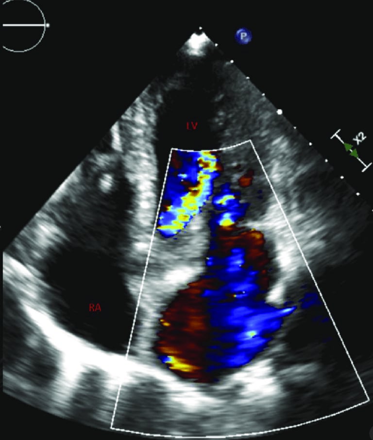
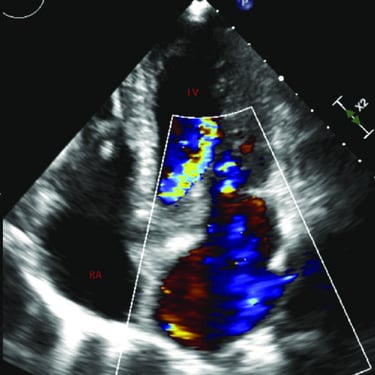
Transthoracic Echocardiogram
Stress ECG
Holter ECG
Tilt Test
Holter Blood Pressure
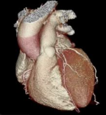
Cardiac CT
Cardiac MRI
An ECG measures electrical activity in the heart, identifying heart rhythm abnormalities and other heart conditions
An echocardiogram is an ultrasound scan that uses sound waves to create detailed images of the heart, assessing its structure, function, and blood flow to diagnose various cardiac conditions.
A treadmill ECG, or exercise stress test, records the heart’s electrical activity during physical exertion to assess its response to stress, helping diagnose conditions like coronary artery disease and exercise-induced arrhythmias.
A Holter blood pressure monitor is a portable device that records blood pressure over 24 hours to detect fluctuations, assess hypertension control, and identify abnormal patterns that may require treatment adjustments.
A tilt table test assesses causes of unexplained dizziness/fainting by monitoring heart rate and blood pressure while tilting patient to an upright position. It helps diagnosing of conditions as postural hypotension and PoTS.
A portable device that records heart rhythm over an extended period (up to a week), helping detect intermittent heart rhythm problems.
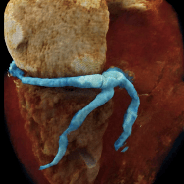
A cardiac CT scan is a non-invasive imaging test that uses X-rays to create detailed 3D pictures of the heart and blood vessels. It helps doctors detect coronary artery disease, calcium buildup, structural heart problems, or congenital abnormalities.
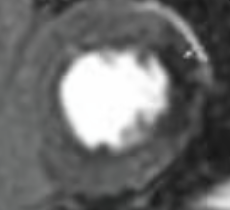
A cardiac MRI scan is a safe and painless test that uses magnetic fields instead of X-rays to produce detailed images of the heart’s structure and function. It helps doctors assess heart muscle health, blood flow, valve function, and any damage after a heart attack. for radiation.
Invasive Procedures
Invasive Angiography
Coronary Angioplasty ± Stenting
Intracoronary Imaging
Microvascular Function & Vasoreactivity Tests
Renal Denervation
Coronary Sinus Reducer
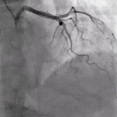
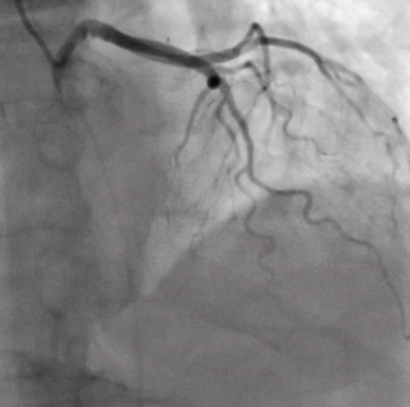
Invasive coronary angiography is a procedure that uses contrast dye and X-rays to visualise the heart’s arteries, assessing for blockages. It is performed under local anaesthesia, typically via wrist access, to guide treatment decisions for coronary artery disease.
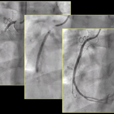
Coronary Angioplasty and stenting are minimally invasive procedures used to treat blocked or narrowed heart vessels. The procedure involves inserting a thin catheter with a balloon into the affected artery, usually via the wrist (radial) or groin (femoral) artery, under local anaesthesia. The balloon is inflated to widen the artery, and a stent (a small metal mesh tube) is placed to keep it open, restoring blood flow. The goal is to relieve chest pain (angina), improve heart function, and reduce the risk of heart attacks.
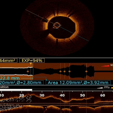
Intracoronary imaging, including Optical Coherence Tomography (OCT) and Intravascular Ultrasound (IVUS), are advanced techniques used during coronary angiography to obtain detailed images of the inside of the heart’s arteries. Performed during invasive angiography or angioplasty, these imaging methods provide high-resolution, real-time views of plaque composition, vessel structure, and stent placement. OCT uses infrared light for microscopic detail, while IVUS employs ultrasound for deeper tissue penetration. These techniques help optimise stent deployment, assess plaque characteristics, and improve precision in coronary interventions, leading to better patient outcomes.
The Coronary Sinus Reducer (CSR) is an hourglass-shaped stent designed for patients with refractory angina. The coronary sinus, a large vein draining blood from the heart, is targeted by the CSR. By implanting the device into the coronary sinus, it narrows the vein, increasing pressure and improving blood flow to ischaemic areas of the heart. The procedure is performed via a catheter-based approach, inserted through a vein in the neck under local anaesthesia. This procedure helps relieve angina symptoms and optimise myocardial perfusion. The CSR offers a non-invasive solution that provides symptom relief and improves quality of life in patients with limited treatment options. It is particularly valuable for those who have failed other interventions, offering significant functional benefits without the need for surgical revascularization.
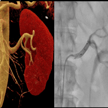
Renal denervation is a minimally invasive procedure designed to treat resistant hypertension, a condition where blood pressure remains high despite at least three medications. It targets the overactive sympathetic nerves surrounding the renal arteries, which contribute to elevated blood pressure.The procedure is performed using a catheter inserted via the femoral or radial artery, which delivers radiofrequency energy or ultrasound waves to disrupt these nerves. This reduces their excessive signalling, leading to a sustained blood pressure reduction. Renal denervation is particularly beneficial for patients with difficult-to-control hypertension, helping lower the risk of heart attack, stroke, kidney disease, and other complications.
Find more Information at oxfordrenaldenervation.com
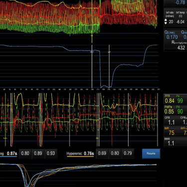
Coronary microvascular function and vasoreactivity tests assess the ability of the small heart vessels to regulate blood flow, helping diagnose conditions like microvascular angina. These tests are performed during invasive coronary angiography by administering medications such as acetylcholine or adenosine through a catheter placed in the coronary artery. These substances induce controlled changes in vessel tone, allowing measurement of microvascular resistance, endothelial function, and vasodilation capacity. The results provide crucial insights into microvascular dysfunction, guiding personalised treatment to improve symptoms and prevent long-term complications.


© 2025. All rights reserved.